Hold down the T key for 3 seconds to activate the audio accessibility mode, at which point you can click the K key to pause and resume audio. Useful for the Check Your Understanding and See Answers.
While the entire surface of the retina contains nerve cells, there is a small portion with a diameter of approximately 0.25 mm where the concentration of cones is greatest. This region, known as the fovea centralis, is the optimal location for the formation of the image. The eye typically rotates in its socket in order to focus images of objects at this location. The distance from the outer surface of the cornea (where the light undergoes most of its refraction) to the central portion of the fovea on the retina is approximately 2.4 cm. Light entering the cornea must produce an image with a distance of 2.4 cm from its outer edge. Unlike a camera, which has the ability to change the distance between the film (the detector) and the lens, the distance between the retina (the detector) and the cornea (the refractor) is fixed. The image distance is unchangeable. Subsequently, the eye must be able to alter the focal length in order to focus images of both nearby and far away objects upon the retinal surface. As the object distance changes, the focal length must be changed in order to keep the image distance constant.
 Accommodation
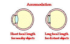
The ability of the eye to adjust its focal length is known as accommodation. Since a nearby object (small dobject) is typically focused at a further distance (large dimage), the eye accommodates by assuming a lens shape that has a shorter focal length. This reduction in focal length will cause more refraction of light and serve to bring the image back closer to the cornea/lens system and upon the retinal surface. So for nearby objects, the ciliary muscles contract and squeeze the lens into a more convex shape. This increase in the curvature of the lens corresponds to a shorter focal length. On the other hand, a distant object (large dobject) is typically focused at a closer distance (small dimage). The eye accommodates by assuming a lens shape that has a longer focal length. So for distant objects the ciliary muscles relax and the lens returns to a flatter shape. This decrease in the curvature of the lens corresponds to a longer focal length. The data table below demonstrates how a changing focal length would be required to maintain a constant image distance of 1.80 cm.
Dependence of f upon dobject
(dimage is fixed at 1.80 cm)
|
Object Distance
|
Focal Length
|
|
0.25 m
|
1.68 cm
|
|
1 m
|
1.77 cm
|
|
3 m
|
1.79 cm
|
|
100 m
|
1.80 cm
|
|
Infinity
|
1.80 cm
|
(The values above have been calculated using the lens equation. The lens equation represents a simplified mathematical model of the eye.)
|
The ability of the eye to accommodate is automatic. Furthermore, it occurs instantaneously. Focus on a far away object and quickly turn your attention to a nearby object; observe that there is no noticeable delay in the ability of the eye to bring the nearby object into focus. Accommodation is a remarkable feat!
The Diopter
The power of a lens is measured by opticians in a unit known as a diopter. A diopter is the reciprocal of the focal length.
diopters = 1/(focal length)
A lens system with a focal length of 1.8 cm (0.018 m) is a 56-diopter lens. A lens system with a focal length of 1.68 cm is a 60-diopter lens. A healthy eye is able to bring both distant objects and nearby objects into focus without the need for corrective lenses. That is, the healthy eye is able to assume both a small and a large focal length; it would have the ability to view objects with a large variation in distance. The maximum variation in the power of the eye is called the Power of Accommodation. If an eye has the ability to assume a focal length of 1.80 cm (56 diopters) to view objects many miles away as well as the ability to assume a 1.68 cm focal length to view an object 0.25 meters away (60 diopters), then its Power of Accommodation would be measured as 4 diopters (60 diopters - 56 diopters).
The healthy eye of a young adult has a Power of Accommodation of approximately 4 diopters. As a person grows older, the Power of Accommodation typically decreases as a person becomes less able to view nearby objects. This failure to view nearby objects leads to the need for corrective lenses. In the next two sections of Lesson 6, we will discuss the two most common defects of the eye - nearsightedness and farsightedness.
Ever wonder what the eye chart in the Doctor's office would look like if you had poor vision? Now you can find out. Use the Imperfect Vision and the Eye Chart widget below to see what the Snellen eye chart would look like with imperfect vision.